Obesity as a Risk Factor for Prostatic Enlargement: A Retrospective Cohort Study in Korea
Article information

Abstract
Purpose
We aimed to evaluate obesity, a risk factor of metabolic syndrome, and its association with prostatic enlargement in a retrospective cohort in Korea.
Methods
Baseline data were obtained from the Korean Genome and Epidemiology Study on Atherosclerosis Risk of Rural Areas in the Korean General Population (KoGES-ARIRANG). Between March 2015 and November 2015, 2,127 male participants of KoGES-ARIRANG were invited to the Korean Prostate Health Council Screening Program, and 602 participants underwent urological examination, including serum prostate specific antigen measurement and transrectal ultrasonography, and completed the International Prostate Symptom Score questionnaire. The data for 571 participants were analyzed, after excluding 31 men who had a history of prostatic disease or testosterone replacement, or had undergone a prior prostatic surgery or procedure.
Results
Among components of metabolic syndrome, waist circumference had a statistically significant linear correlation with incremental increases in prostate volume (B=0.181, P=0.004). Abdominal obesity as determined by anthropometric measures including body mass index (odds ratio [OR], 1.205; 95% confidence interval [CI], 1.088–1.336), waist circumference (OR, 1.073; 95% CI, 1.032–1.115), body fat (OR, 1.126; 95% CI, 1.056–1.202), and visceral fat composition (OR, 1.667; 95% CI, 1.246–2.232) was significantly associated with the presence of high-volume benign prostatic hyperplasia (BPH) (prostate volume≥ 40 mL). Furthermore, the highest quartile of serum leptin (OR, 3.541; 95% CI, 1.103–11.365) and adiponectin levels (OR, 0.315; 95% CI, 0.102–0.971) were significantly correlated with high-volume BPH compared to the lowest quartile of levels.
Conclusions
Abdominal obesity and serum leptin level are positively associated with prostate growth, whereas serum adiponectin level is inversely associated with the presence of prostatic enlargement.
INTRODUCTION
Benign prostatic hyperplasia (BPH) is one of the most common conditions in elderly men [1]. Although not fatal, BPH leads to troublesome lower urinary tract symptoms (LUTS) that can diminish quality of life (QoL) and results in economic and social consequences [2]. However, the etiology and pathogenesis of BPH remain unclear. Previous studies have shown that BPH is associated with sex steroid hormone levels and has a genetic predisposition [3]. Recently, attention has been focused on diet, exercise, lifestyle, and metabolic disturbances as risk factors for BPH, in addition to well-established factors, such as age and testosterone level [4,5].
Since the concept of syndrome X was introduced as comprising risk factors for atherosclerosis in 1987, metabolic syndrome (MetS) has had wide-ranging effects on modern medical care [6]. MetS is a combination of medical disorders, including obesity, impaired glucose metabolism, hypertension (HTN), hypertriglyceridemia, and low high density lipoprotein cholesterol; these affect the occurrence of cardiovascular diseases (CV) and diabetes mellitus (DM) type 2 [4,5]. Although risk factors for MetS have been identified, insulin resistance is likely to be a significant link between the components of MetS. Moreover, it seems clear that obesity results in the development of insulin resistance and has triggered the escalating incidence of MetS [7].
Obesity is a medical condition in which excess body fat has accumulated and, particularly in the Western world, is a well-known problem in society. However, the prevalence of obesity in developing countries is increasing toward that currently seen in developed countries. Thus, the increase in obesity rates is becoming a serious health problem, leading the American Medical Association to classify obesity as a disease in 2013 [8]. Accordingly, obesity is projected to be the most common modifiable factor among preventable causes of death in the world [8].
Interestingly, a few large cohort studies found that obesity was correlated with the risk of symptomatic BPH [9,10]. In addition, a recent study found that serum adipokines, which are metabolically active hormones secreted by excess adipose tissue, were associated with the risk of symptomatic BPH [11]. Therefore, we evaluated obesity-related factors and their association with BPH in a population-based cohort in Korea.
MATERIALS AND METHODS
Study Population and Design
Baseline data for the study were obtained from the Korean Genome and Epidemiology Study on Atherosclerosis Risk of Rural Areas in the Korean General Population (KoGES-ARIRANG), a population-based prospective cohort study which assessed the prevalence and incidence of and risk factors for chronic degenerative disorders such as HTN, DM, CV, and MetS in rural areas of Wonju and Pyeongchang in South Korea. The baseline survey was conducted from November 2005 to January 2008 and included 5,178 adults (2,127 men and 3,051 women) aged 40–70 years. Between March 2015 and November 2015, all male participants of KoGES-ARIRANG were invited to participate in the Korean Prostate Health Council Screening Program, which provided a free medical examination to men in the general community. Six hundred two participants of the KoGES-ARIRANG attended the screening program. Participants who had a history of prostatic disease or had undergone prior prostatic surgery or procedure in the baseline KoGES-ARIRANG survey were excluded. The participants on testosterone replacement were also excluded. The remaining 571 participants were included in the analysis. This study was approved by the Institutional Review Board of the Yonsei University Wonju College of Medicine (YWMR-15-9-017).
Measurements of Prostate-Specific Characteristics
All participants underwent a urological examination, including measurement of serum prostate-specific antigen (PSA) level and prostate volume (PV) and completion of an International Prostate Symptom Score (IPSS) questionnaire. PSA level was assayed with commercially available kits using the chemiluminescence method (Siemens, Malvern, PA, USA). PV was determined using transrectal ultrasonography, which was performed using a 7.5-MHz rectal probe (Aloka, Tokyo, Japan); PV was then calculated using the ellipsoid formula {PV=π/6×[width (cm)×thickness (cm)×length (cm)]} [12]. The IPSS questionnaire is composed of three domains related to storage symptoms (frequency, urgency, and nocturia), four domains related to voiding symptoms (hesitancy, weak stream, intermittence, and incomplete emptying), and one domain related to QoL. Each IPSS domain uses a 6-point scale ranging from 0 (none) to 5 (5 or more). The total score for symptoms was defined as the sum of scores for seven domains: the total score ranges from 0 to 35, with a higher score indicating a higher severity of symptoms [3]. Uroflowmetry data (Medtronics, Minneapolis, MN, USA) were collected to calculate maximal flow rate (Qmax). Qmax <10 mL/sec reflects the presence of bladder outlet obstruction, such as that which occurs in BPH [3].
Measurements of Demographic and Metabolic Characteristics
At the baseline KoGES-ARIRANG examination, study participants completed a standardized medical history and health questionnaire. Baseline information on smoking was collected using a self-reported questionnaire (yes or no). Height, body weight, waist circumference (WC), and blood pressure (BP) were determined according to international standards. Body mass index (BMI) was calculated as weight (kg)/height (m2). Body fat and visceral fat composition were determined using a multifrequency impedance body composition analyzer with 8-point tactile electrodes (Jawon Medical Co. Ltd., Kyungsan, Korea). Venous blood samples were drawn from each participant after an 8-hour period of fasting. Fasting glucose and insulin were determined using a glucose oxidase-based assay and double antibody radioimmunoassay (Biosource Europe SA, Nivelles, Belgium), respectively. Serum concentrations of total cholesterol, low-density lipoprotein (LDL) cholesterol, high-density lipoprotein cholesterol, and triglycerides were determined using enzymatic methods (Advia 1650, Siemens, Tarrytown, NY, USA). Hemoglobin A1c was determined by high-performance liquid chromatography (Variant II, Bio-Rad, Hercules, CA, USA). Homeostasis model assessment of insulin resistance (HOMA-IR) values were calculated using the following formula: fasting plasma glucose (mg/dL) ×fasting insulin (mIU/mL)/22.5 [13]. High sensitivity C-reactive protein (CRP) was measured using the Denka Seiken (Tokyo, Japan) assay. The serum concentrations of adiponectin were measured using a radioimmunoassay (LINCO Research, St. Charles, MO, USA), with intra-assay and interassay coefficients of variation ranging between 2.9% and 6.6%.
Statistical Analysis
Baseline characteristics of participants were assessed using the one-way analysis of variance and chi squared test according to the properties of the variable. Linear regression analysis was used to evaluate the relationship between MetS risk factors and PV. The univariate model was adjusted for age, smoking history, LDL, and PSA; each metabolic component was evaluated individually. The result of multivariate analysis was the combined effect of all factors after adjustment for age, smoking history, LDL, and PSA. Logistic regression analysis was conducted to evaluate the association of obesity related factors, including BMI, WC, body fat and visceral fat composition, and adipokines with prostatic enlargement. Model I was unadjusted and model II was adjusted for age, smoking history, LDL, and PSA. All statistical analyses were performed using IBM SPSS Statistics ver. 21.0 (IBM Co., Armonk, NY, USA), and a two-tailed P-value of <0.05 was considered statistically significant.
RESULTS
Of 571 participants, the mean age was 58.53±7.01 years (range, 40–72 years), and the mean serum PSA level was 1.41±2.48 mg/mL (range, 0.02–40.10 mg/mL). The mean PV was 27.82±11.08 mL (range, 10.00–128.30 mL), and mean IPSS was 11.28±7.50 (range, 0–35). The mean Qmax rate was 12.43 ±7.33 mL/sec (range, 0.70–42.60 mL/sec). The mean PSA level increased significantly in accordance with PV, but other prostate-specific characteristics were not significantly different (Table 1).

Baseline characteristics of participants according to prostate volume
Anthropometric and Metabolic Characteristics (Table 1)
All participants were stratified into three groups according to PV. The mean BMI of all participants was 24.58±2.85 kg/m2 (range, 15.73–36.41 kg/m2). The mean body fat and visceral fat mass were 16.57±4.58 kg (range, 2.30–33.70 kg) and 2.54±0.93 kg (range, 0.20–6.80 kg), respectively. The mean WC was 87.16 ±7.66 cm (range, 62.00–118.00 cm). The mean serum leptin level was 2.76±1.99 μg/L (range, 0.39–14.68 μg/L). Obesity-related anthropometric characteristics, such as BMI, body fat composition, visceral fat composition, and WC were significantly different between groups (P=0.003, P=0.007, P=0.008, and P=0.008, respectively). In addition, leptin, a well-known adipokine, was significantly different among the 3 groups (P=V0.014). However, other metabolic characteristics reflecting insulin resistance, dyslipidemia, and BP were not significantly different. Adiponectin, which has a variety of protective properties against metabolic disturbances, also did not demonstrate a statistically significant difference between groups.
Metabolic Risk Factors and PV (Table 2)
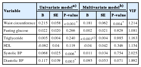
Correlation between metabolic components and prostate volume
The univariate model showed that WC and BP were significantly correlated with PV (WC: B=0.215, P<0.001; systolic BP: B=0.068, P=0.006; diastolic BP: B=0.117, P=0.003). On multivariate analysis for the combined effect of all factors after adjustment for age, smoking history, LDL, and PSA, only WC had a statistically significant linear correlation with incremental increases in PV (B=0.181, P=0.004). All variance inflation factors were <10.
Obesity Related Factors and Prostatic Enlargement (Table 3)

Multivariate logistic analysis for obesity related risk factors associated with development of benign prostatic hyperplasia
As shown in Tables 1 and 2, obesity was significantly correlated with PV. To confirm the association between obesity related factors and prostatic enlargement, logistic analysis was performed. PV between 30 and 39 mL was defined as low-volume BPH, and PV≥40 mL was defined as high-volume BPH. For this analysis, we also divided leptin and adiponectin levels into quartiles.
On the unadjusted model (model I), BMI, WC, body fat composition, and visceral fat composition were significantly associated with the presence of high-volume BPH (BMI: odds ratio [OR], 1.115; WC: OR, 1.051; body fat: OR, 1.088; visceral fat: OR, 1.463). However, no obesity-related factors were significantly related to the presence of low-volume BPH. In addition, increasing quartiles of adipokines were not significantly associated with high-volume BPH.
On the adjusted model (model II, which controlled for age, smoking history, LDL, and PSA), BMI, WC, body fat composition, and visceral fat composition were also significantly associated with the incidence of high-volume BPH (BMI: OR, 1.205; WC: OR, 1.073; body fat: OR, 1.126; visceral fat: OR, 1.667). Furthermore, compared to the lowest leptin level quartile, the highest quartile was significantly associated with high-volume BPH (OR, 3.541). In contrast, compared to the lowest adiponectin level quartile, the highest quartile displayed a significantly lower probability of high-volume BPH (OR, 0.315). No obesity-related factors were statistically significantly associated with the presence of low-volume BPH.
DISCUSSION
Growing evidence suggests that obesity, which is a component of MetS, has been associated with an increased risk of several common adult cancers [14]. From a urological standpoint, an association between obesity and an increased cancer risk has been reported in renal cell carcinoma [15]. Additionally, epidemiologic studies and a meta-analysis have shown that obesity has been consistently associated with poorer outcome and a higher-grade disease in prostate cancer [16-18]. Aside from urinary cancer, a recent review found an association between obesity and nonmalignant urological disease, such as BPH [19,20]. Moreover, in a prospective cohort, each 1-kg/m2 increase in BMI was associated with a 0.41-cm3 increase in PV [21]. In the Prostate Cancer Prevention Trial, the incidence of BPH, which was defined as severe urinary symptoms as determined by the IPSS or the initiation of medical or surgical therapy, increased 10% for each 0.05 increase in waist-to-hip ratio [10]. This is consistent with the present findings, which showed that obesity, as determined by anthropometric measures, is positively associated with PV. Furthermore, WC was a significant factor in predicting the presence of high-volume BPH.
Although the molecular mechanisms and the pathophysiology by which obesity may promote BPH are not fully understood, the likely biologic mechanism seems to be insulin resistance [4]. Briefly, the chronic elevation of insulin is associated with the increased availability of insulin-like growth factor-1 (IGF-1). Insulin has been suggested to stimulate tumorigenesis by inducing IGF-1 synthesis and activating insulin and IGF-1 receptors, which are often overexpressed in cancer cells. Moreover, insulin and IGF-1 interactions with downstream signaling pathways may impact the growth of hormonally driven tumors, such as prostate cancer; they may also stimulate BPH [11,22]. In addition, insulin may influence the transcription of genes involved in sex hormone metabolism; these genes alter the androgen/estrogen ratio and circulating level of sex hormonebinding globulin. Such alterations in the testosterone to estrogen ratio in prostate tissue may contribute to BPH development [21,23]. Another potential explanation involves chronic inflammation and oxidative stress. Increased BMI is associated with adipocyte hypertrophy and death, which cause excessive cytokine production and leukocyte recruitment. These inflammatory changes in the tissue may provide a pro-neoplastic microenvironment [22,24]. However, in the present study, serum insulin level and HOMA-IR, which are indicators of insulin resistance, were not significantly different according to PV, although they were higher in patients with high-volume BPH than in those without BPH. Furthermore, the number of leukocytes and serum CRP level, which are markers of inflammation, were not statistically higher in the participants with prostatic enlargement. The reasons for this finding are unclear and should be further examined in future studies. Finally, adipokines, such as leptin and adiponectin, which maintain metabolic homeostasis and balance cell proliferation and apoptosis, may play a key role in BPH pathogenesis due to their vital role in regulating body weight [25]. Leptin promotes cell proliferation through alteration of cell cycle checkpoints and upregulation of specific genes, advancing cell cycles from G1 to S phase. Leptin also has both proinflammatory and angiogenic effects, which may result in carcinogenesis [22,26]. Adiponectin has properties in apoptosis and metabolism of glucose and fatty acids [25]. In vitro, adiponectin suppresses dihydrotestosterone-stimulated cell proliferation, which is involved in the regulation of prostatic growth [27]. In addition, adiponectin enhances insulin sensitivity by inhibiting both the expression of hepatic gluconeogenic enzymes and endogenous production of glucose [28]. This suggests that adiponectin may indirectly affect prostatic growth via insulin sensitivity. Previous studies showed that altered levels of adipokines, particularly increased leptin and decreased adiponectin, are commonly associated with obesity [29]. Schenk et al. [25] reported an association of high adiponectin concentrations with a reduced risk of BPH. However, they found that leptin and CRP were not associated with BPH risk. The current study showed that the highest leptin level quartile was significantly correlated with high-volume BPH and the highest adiponectin level quartile was inversely correlated with high-volume BPH. These findings suggest that the balance between these two adipokines may be associated with obesity-induced prostatic growth.
The present study has several limitations. Firstly, although we used the data of KoGES-ARIRANG, we retrospectively conducted a cross-sectional study. Secondly, there may be selection bias, as the study participants participated in KoGES-ARIRANG and were further recruited to participate in the Korean Prostate Health Council Screening Program. Thus, many of these participants may already be proactively concerned with their health and have a good doctor-patient relationship. Thirdly, definitions regarding the appropriate BPH cutoff points are controversial. However, we used PV as the only measure for diagnosis of BPH to focus on the relationship between obesity and PV. Finally, the participants with comorbidities that can affect metabolic profile parameters were included in this study, although there were no statistically significant differences between groups. Accordingly, further longitudinal studies are needed to elucidate the relationship between MetS and BPH.
Nonetheless, we believe that obesity management and prevention may be a novel target for the prevention of BPH. Previous literature has indicated that the detrimental effect of obesity is reversible and long-term weight loss was shown to have a significant effect on bladder pressure and urinary incontinence [19,30]. Moreover, a meta-analysis of 11 published studies concluded that moderate to vigorous physical activity was associated with a 25% lower risk for BPH or LUTS [31]. This indicates that physical activity and lifestyle modification may have a therapeutic effect in men with BPH. These data show that many of the metabolic disturbances associated with cardiovascular disease and their modulating lifestyle factors may be associated with BPH onset and progression. Therefore, urologists should consider the effect of obesity on urological health as well as overall public health.
Notes
Research Ethics
This study was approved by the Institutional Review Board of the Yonsei University Wonju College of Medicine (YWMR-15-9-017).
Conflict of Interest
No potential conflict of interest relevant to this article was reported.
Acknowledgements
We are grateful to all members of the Korean Prostate Health Council Screening Program in the preparation of this article.