INTRODUCTION
Overactive bladder (OAB) is defined as the presence of urinary urgency, usually accompanied by frequency and nocturia, with or without urgent incontinence, in the absence of urinary tract infection and other urethrovesical dysfunction [1]. OAB is a micturition-related symptom complex; however, it affects not only the discomfort but also the quality of life for all ages. More than 16% of men and women over 40-year-old suffer from OAB [2]. Continence and urination is related to the balance of the relaxation and the contraction of the detrusor and sphincter muscles. Therefore, there is no spastic detrusor muscle contraction during the storage phase. In OAB patients, however, uninhibited spastic detrusor muscle contractions occur and result in sustained high bladder pressure, causing urinary urgency or urgency incontinence [3]. OAB patients experience depression and complain of sleep disturbances, and these effects disturb quality of life [2].
Antimuscarinic agents reduce bladder contraction frequency and pressure, so these drugs are currently used for the treatment of OAB. However, side effects of antimuscarinic agents, such as dry mouth, impaired cognitive function, constipation, and blurred vision, lead to low patient compliance [2,4].
Alpha 1-adrenergic receptor (α1-AR) antagonists are the most well-known drugs to improve lower urinary tract symptoms (LUTS), and α1-AR antagonists have been used to treat micturition symptoms of OAB [5]. Each α1-AR antagonist has a unique affinity with or selectivity toward the AR subtypes, showing different actions and side effects [6]. Combined usage of α1-AR antagonists without any concurrent proof or investigation might cause adverse effects. In the AR antagonist era, in contrast, the combination therapies might function complementarily with more efficacy and safety, because each α1-AR antagonist has its own characteristics in receptor selectivity, central nervous system permeability, and risk of side effects. Therefore, there is a need to evaluate the synergistic effects and/or side effects in the combination therapy of α1-AR antagonists.
We investigated the efficacy of add-on therapy of α1-AR antagonists on the OAB animal model using urodynamic techniques and neurophysiologic methods. Cystometry was performed to evaluate contraction pressure and time, and immunohistochemical staining was conducted to determine c-Fos and nerve growth factor (NGF) expressions in the central micturition centers (medial preoptic nucleus [MPA], ventrolateral periaqueductal gray [vlPAG], pontine micturition center [PMC], and spinal cord L4–L5).
MATERIALS AND METHODS
Animal Treatments
Adult female Sprague-Dawley rats, weighing 230 ±10 g (10 weeks old), were used for these experiments. The guidelines of the Institutional Care and Use Committee of Kyung Hee University were followed during all laboratory procedures (KHUASP [SE]-14-047), and all experiments were performed in accordance with the guiding principles for the care and use of animals approved by the Council of the National Institutes of Health Guide for the Care and Use of Laboratory Animals. All rats were randomly divided into the following 5 groups (n=10 in each group): (1) control group, (2) OAB-induction group (OAB), (3) OAB-induction and tamsulosin monotherapy group (OAB-Tam), (4) OAB-induction and naftopidil monotherapy group (OAB-Naf), and (5) OAB-induction and tamsulosin-naftopidil combination therapy group (OAB-Combi). Each drug was administered with reference to the recommended daily allowance in each group: tamsulosin (0.2 mg/kg, Harunal, Astellas Pharma Inc., Tokyo, Japan), naftopidil (75 mg/kg, Flivas, Asahi Kasei Pharma, Tokyo, Japan), and combination (0.2 mg/kg tamsulosin+75 mg/kg naftopidil). The rat in the drug-treated groups received each drug by oral administration once a day for 14 days. For the rats in the control group and in the OAB-induction group, distilled water of the same volume was orally administrated for the same duration.
Induction of OAB
The OAB model was induced by the previously described method [4]. For the induction of the OAB model, 75 mg/kg of cyclophosphamide (Sigma Chemical Co., St. Louis, MO, USA) was intraperitoneally injected every third day for 10 days. The rats in the control group received intraperitoneally volumematched saline.
Cystometry
The contraction pressure and time in the cystometry were evaluated using the previously described method 14 days after OAB induction [4]. After a transperitoneal incision, a polyethylene catheter (PE50) was positioned into the bladder under the anesthesia with an intraperitoneal Zoletil 50 (10 mg/kg; Vibac Laboratories, Carros, France) injection. Bladder pressure was evaluated by connection to a syringe pump (Havard Apparatus, Holliston, MA, USA) and pressure transducer (Havard Apparatus) through a 3-way stopcock to infuse saline into the bladder and to record intravesical pressure simultaneously. After bladder emptying, a pressure-flow study was performed with a 0.5 mL/sec saline infusion. The contraction pressure and contraction time of the bladder were recorded using Labscribe (iWork System Inc., Dover, NH, USA).
Tissue Preparation
The rats were sacrificed immediately after the cystometry evaluation. After administration of anesthesia with intraperitoneal injection of Zoletil 50 (Vibac Laboratories), a 50mM phosphate-buffered saline transcardial perfusion was performed. The fixation was done with a fresh solution consisting of 4% paraformaldehyde in a 100mM phosphate buffer (pH, 7.4). After that, it remained overnight with the same fixative after the brain and spinal cord dissection, and then it was transferred to 30% sucrose solution for cryoprotection. For the immunohistochemical staining, the brain was sliced with the coronal sectioned at 40 μm thick using a cryostat (Leica, Nussloch, Germany). Each location of the central micturition center was designated in accordance with the previous method [4]. The MPA, vlPAG, and PMC were designated ranges –0.26 to 0.80 mm, –7.64 to 8.00 mm, and –9.68 to –9.80 mm from Bregma, respectively. To get the micturition center in the spinal cord, we harvested the L4–L5 regions. On average, 8 slices were collectable in each region from each rat.
Immunohistochemistry for c-Fos and NGF
Immunohistochemistry was conducted to evaluate the expressions of c-Fos and NGF in the central micturition centers (MPA, vlPAG, PMC, and spinal cord L4–L5) in accordance with the previously described method [7]. Free-floating slices were cultivated with rabbit anti-c-Fos antibody and mouse antiNGF antibody (1:1,000; Santa Cruz Biotechnology, Santa Cruz, CA, USA) at a dilution of 1:1,000 overnight. Further incubation was performed with biotinylated anti-rabbit secondary antibody for c-Fos and anti-mouse secondary antibody for NGF (1:200; Vector Laboratories, Burlingame, CA, USA). Then, the slices were cultivated with avidin-biotin-peroxidase complex (Vector Laboratories) for 1 hour at room temperature. Following the incubation in the reaction mixture consisting of 0.03% diaminobenzidine tetrahydrochloride and 0.03% hydrogen peroxide, the sections were put onto gelatin-coated slides. After air-drying overnight at room temperature, the cover-glasses were covered up using Permount (Fisher Scientific, New Jersey, NJ, USA).
Data Analysis
The numbers of c-Fos-stained and NGF-stained cells were counted hemilaterally under a light microscope (Olympus, Tokyo, Japan). Then an Image-Pro Plus computer-assisted image analysis system (Media Cyberbetics Inc., Silver Spring, MD, USA), which was assembled with light microscope (Olympus), was used to measure the neuronal voiding centers of each slide. For statistical analysis, a one-way analysis of variance and Duncan post hoc test was used. All results were written as the mean± standard error of the mean, and P<0.05 was considered to be statically significant.
RESULTS
Effect of α1-AR Antagonists on Bladder Function
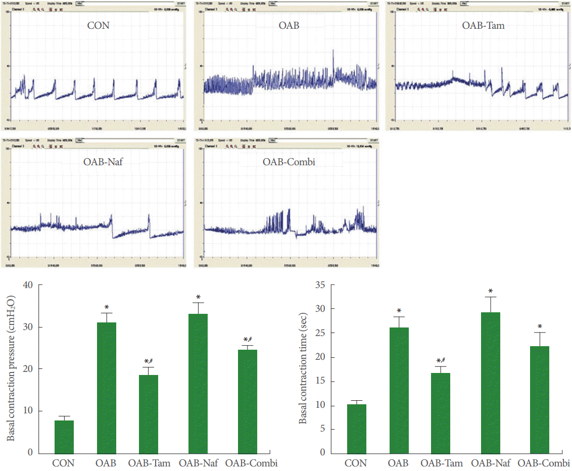
The bladder contraction pressure and time are presented in Fig. 1. The bladder contraction pressure was 8.02±0.80 cm H2O in the control group, 31.16±2.04 cm H2O in the OAB group, 18.64±1.66 cm H2O in the OAB-Tam group, 33.16 ±2.38 cm H2O in the OAB-Naf group, and 24.16 ±1.08 cm H2O in the OAB-Combi group. The contraction time was 10.33±0.82 seconds in the control group, 26.11±2.15 seconds in the OAB group, 16.75±1.29 seconds in the OAB-Tam group, 29.12±3.07 seconds in the OAB--Naf group, and 22.16±2.75 seconds in the OAB-Combi group.
In the present results, contraction pressure and time were increased by induction of OAB, whereas, tamsulosin and combination treatment decreased the OAB-induced contraction pressure.
Effects of α1-AR Antagonists on c-Fos Expressions in the Central Micturition Centers
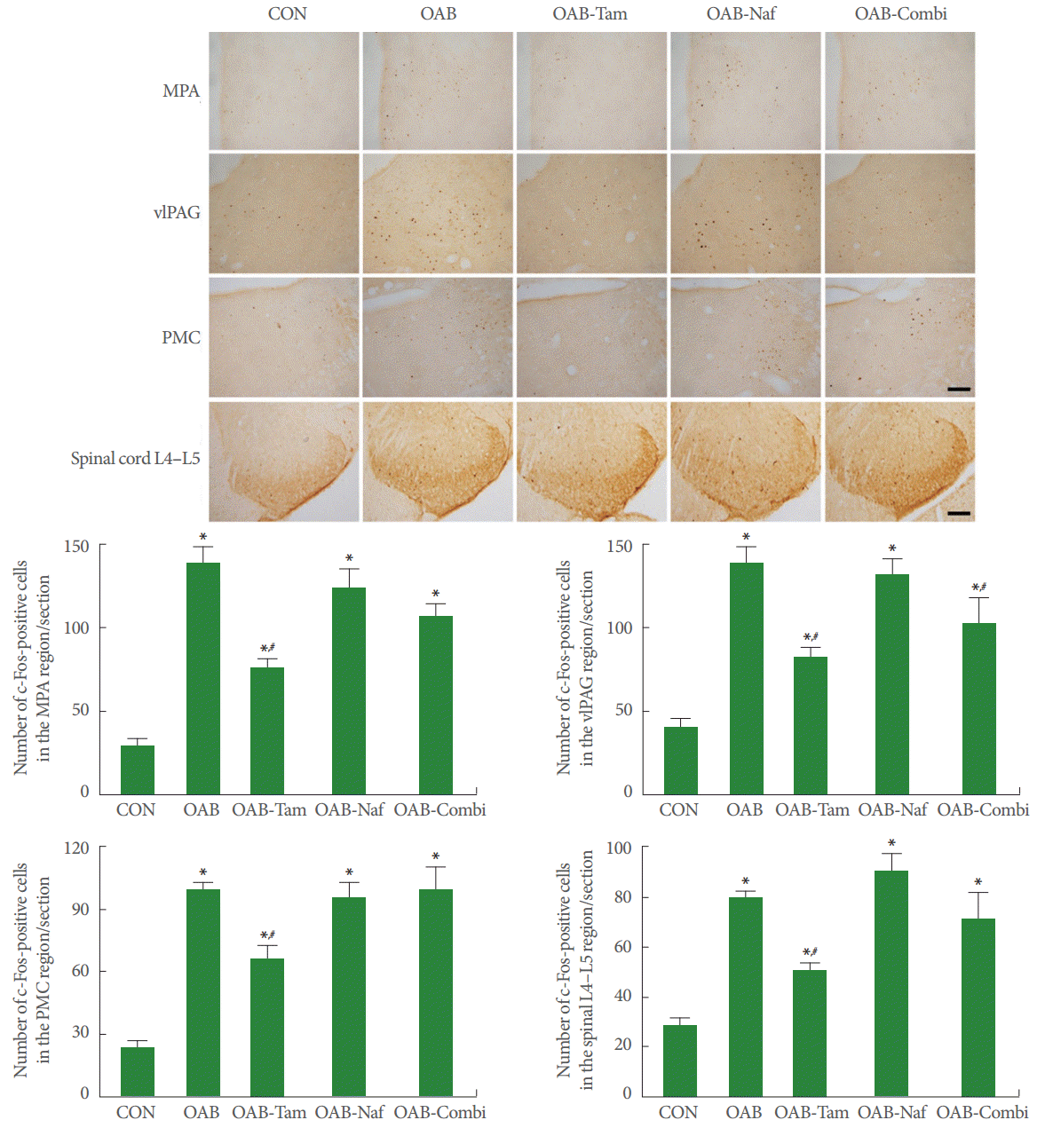
The c-Fos expressions in the central micturition centers (MPA, vlPAG, PMC, L4-L5) are presented in Fig. 2. In the MPA, the c-Fos-positive cell count was 29.32±4.01/section in the control group, 139.36 ±7.96/section in the OAB group, 75.48 ±5.11/section in the OAB-Tam group, 123.62±11.15/section in the OAB-Naf group, and 105 ±7.99/section in the OAB-Combi group.
In the vlPAG, the c-Fos-positive cell count was 39.15±5.59/section in the control group, 136.25±11.59/section in the OAB group, 81.60±5.18/section in the OAB-Tam group, 130.03±9.11/section in the OAB-Naf group, and 101.13±14.31/section in the OAB-Combi group.
In the PMC, the c-Fos-positive cell count was 22.12±3.87/section in the control group, 99.16 ±3.04/section in the OAB group, 65.79±6.13/section in the OAB-Tam group, 94.96±7.17/section in the OAB-Naf group, and 98.13±11.01/section in the OAB-Combi group.
In the L4–L5, the c-Fos-positive cell count was 29.19±2.11/section in the control group, 79.89±2.19/section in the OAB group, 50.11±3.01/section in the OAB-Tam group, 89.39±7.11/section in the OAB-Naf group, and 70.39±10.60/section in the OAB-Combi group.
In the present results, c-Fos expressions in the central micturition centers were increased by induction of OAB. Increased c-Fos expressions were suppressed by tamsulosin, however, naftopidil and combination treatment did not suppress c-Fos expressions.
Effects of α1-AR Antagonists on NGF Expressions in the Central Micturition Centers
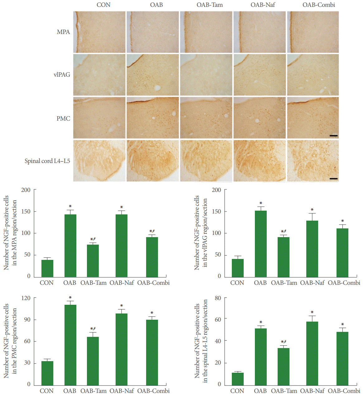
The NGF expressions in the central micturition center (MPA, vlPAG, PMC, L4–L5) are presented in Fig. 3. In the MPA, the NGF-positive cell count was 39.38±5.01/section in the control group, 141.59±10.33/section in the OAB group, 74.19±3.97/section in the OAB-Tam group, 139.99 ±9.28/section in the OAB-Naf group, and 89.16±4.18/section in the OAB-Combi group.
In the vlPAG, the NGF-positive cell count was 41.28±5.96/section in the control group, 150.19±8.39/section in the OAB group, 90.39±4.19/section in the OAB-Tam group, 129.19±15.97/section in the OAB-Naf group, and 109.18±8.19/section in the OAB-Combi group.
In the PMC, the NGF-positive cell count was 33.19±2.09/section in the control group, 109.60±4.50/section in the OAB group, 66.01±5.10/section in the OAB-Tam group, 98.10±4.98/section in the OAB-Naf group, and 89.77±2.70/section in the OAB-Combi group.
In the L4–L5, the NGF-positive cell count was 11.20±1.40/section in the control group, 51.01±2.50/section in the OAB group, 33.20±2.11/section in the OAB-Tam group, 56.99±5.17/section in the OAB-Naf group, and 47.19±4.19/section in the OAB-Combi group.
In the present results, NGF expressions in the central micturition centers were increased by induction of OAB. Increased NGF expressions were suppressed by tamsulosin; however, naftopidil and combination treatment did not suppress NGF expressions.
DISCUSSION
Normal bladder filling and storage processes require cooperation among volume, pressure, and appropriate sensation. If bladder volume increases without an increment of intravesical pressure, urinary urge sensation occurs. The urethral sphincter should simultaneously retain closed and be without involuntary bladder contraction [8]. On the contrary, such an increase in the basal contraction pressure and time and uninhibited bladder contraction are found in OAB [4].
In the present results, contraction pressure and time were increased by cyclophosphamide administration, showing that OAB was induced by repeated cyclophosphamide injections. Furthermore, increased expressions of c-Fos and NGF in the central micturition centers were caused by repeated cyclophosphamide injections, representing neuronal activation.
For treatment of voiding and storage symptoms of OAB, α1-antagonists are able to use [9]. Tamsulosin has an α1A-AR subtype dominant affinity than the ;1;1D-AR subtype, and tamsulosin exerts distinct actions on the prostate, external sphincter, and urethra. Meanwhile, naftopidil has a 3-fold affinity for the α1D-AR subtype rather than the α1A-AR subtype, and naftopidil shows different characteristics on LUTS, because major α1DAR exists in the bladder neck [10]. Both α1-AR subtype antagonists alleviate OAB-related symptoms, such as frequent micturition and urinary urge sensations [11]. Frequency, urgency, nocturia, and urgency incontinence were decreased by α1-AR antagonists in clinical research [12]. Furthermore, α1-AR antagonists increase bladder capacity and decrease frequency [4,13]. Each α1-AR antagonist has its own unique properties because of the differences in affinity and the degree of the effects on the central nervous system [14]. In this study, the supplementary effects on efficacy or side effects in the combination therapy of α1-AR antagonists in relation with central micturition centers were evaluated.
In the present results, increased contraction pressure and time induced by cyclophosphamide injection was suppressed by tamsulosin monotherapy. However, naftopidil monotherapy showed no significant effect on contraction pressure and time. Moreover, combination therapy showed a less significant effect on contraction pressure and time compared to the tamsulosin monotherapy. These controversial phenomena might be ascribed to the antagonistic action of naftopidil on the tamsulosin.
Voiding function is controlled by central micturition centers. Central micturition centers, including the pons and PMC, are implicated in an OAB [15]. Continence with low bladder pressure during much of daily life is acquired by simultaneous excitation of sympathetic motor neurons and suppression of the parasympathetic motor neurons, and it can be achieved by the excitation of α1-AR during the storage phase [16]. During micturition, suppression of the sympathetic motor neurons and activation of the parasympathetic motor neurons occur. PMC neurons directly activate the parasympathetic preganglionic motor neurons causing bladder contraction and sustained relaxation of the urethral sphincter [15,17]. vlPAG is a central region in the controlling micturition through both afferent and efferent pathways. The efferent inhibitory signal passes through the periaqueductal gray (PAG) to the pons, and this excessive inhibitory signal triggers the reflex to the PMC, which results in urethral sphincter relaxation [18]. Activation of PMC neurons initiates urethral sphincter relaxation and detrusor muscle contraction, resulting in urination [19]. The PAG-PMC connection is the major part that controls micturition. The MPA in the hypothalamus sends projections directly to the PMC, except for the vlPAG [20]. vlPAG emits the efferent signal to continue or to stop the inhibition of PMC, suggesting that voiding is under the control of brain areas, such as the prefrontal cortex, the anterior circulate cortex, the insula, and the hypothalamus [18]. PAG activity increases as the bladder volume increases, which means that vlPAG performs an integrating function of the somatic, autonomic, and sensory components of the micturition reflex [21-23].
In the present results, tamsulosin showed an inhibitory effect on cyclophosphamide-induced enhancement of c-Fos and NGF expressions in the central micturition centers. These results suggest that c-Fos and NGF in the voiding centers are implicated in the modulation of micturition and that tamsulosin inhibits neuronal activation in the central micturition centers of OAB rats. However, naftopidil monotherapy and combination therapy exerted no significant effect on the c-Fos and NGF expressions in the central micturition centers of OAB rats. From the present results, we suggest that tamsulosin exerts neuromodulatory effect on voiding centers; in contrast, naftopidil has not such an effect.
Through this experiment, tamsulosin showed the most prominent efficacy for the treatment of OAB compared to the naftopidil or combination. For OAB, a combination of tamsulosin with naftopidil showed no synergistic effects; however, the possibility that a combination of other AR antagonists might improve efficacy on OAB still exists. For example, nonselective AR antagonist alfuzosin causes hypotension without ejaculation problems; on the other hand, tamsulosin causes ejaculation problems without hypotension [24,25]. Meanwhile naftopidil shows minimal effects on the cardiovascular system and ejaculatory function [26]. Further studies of add-on therapy to maximize the treatment efficacy or minimize the side effects might provide an opportunity to find a new modality.