
INTRODUCTION
Urodynamic studies have been used as a functional tool to quantitatively evaluate symptoms of lower urinary tract (LUT) dysfunction and to identify the underlying causes through the measurement of relevant physiologic parameters [1]. It is essential for describing the status of the LUT and is highly recommended in patients with neurogenic LUT dysfunction [2]. Quality control plays a key role in ensuring reliable and accurate urodynamic data. Whether in real-time or a retrospective evaluation, typical value ranges (TVRs) and typical signal patterns (TSPs) are very efficient tools for performing quality control quantitatively and qualitatively. The initial stage of urodynamic studies is crucial because TVRs for signal patterns that are perfect in terms of physiological values usually remain present throughout the investigation [3]. Evaluating the TVRs and TSPs with regard to the initial resting pressure and cough test are crucial quality control steps at the beginning of the urodynamic study. The TVRs of the initial resting pressure can be used to confirm whether the equipment has been set up properly, while the TSPs of minor dynamic changes and cough before filling can be used to check if the signal transmission is functioning properly. The TSPs of the cough signal have been identified in real-time urodynamics [4]. It has been claimed that intravesical pressure (Pves) and abdominal pressure (Pabd) traces show the same fine structures and equivalent major changes, while detrusor pressure (Pdet) does not change significantly during cough tests. Sullivan et al. [5] identified cough signal quality by comparing the measured height of the cough spikes on Pabd and Pves tracings, stating that a good cough signal has a smaller spike that is 70%ŌĆō100% the size of the larger spike. This is a somewhat arbitrary classification of the cough signal. As a tool for quality control, coughing should be quantified to the standards used by urodynamic testing centers. Hogan et al. [6] suggested that transmitted cough amplitudes should have a minimum peak height of 15 cm H2O above the resting pressure; however, the process of implementing such a standard has not been clarified. The aim of this study was to review the consecutive urodynamic traces obtained from neurogenic bladder patients at our center in 2014 to establish the TVRs of the initial cough (cough before bladder filling) signal and to analyze the TSPs of the initial cough.
MATERIALS AND METHODS
We retrospectively reviewed 539 urodynamic traces from patients (366 male patients and 173 female patients) who received treatment in our center in 2014. Their mean age was 36┬▒17 years. All patients had a known diagnose of neurogenic bladder, due to traumatic spinal cord injury (347), myelomeningocele (55), spinal surgery (40), and other conditions (Table 1). Of these patients, 443 underwent urodynamic testing in the supine position, while 96 patients underwent testing in the sitting position. All measurements were obtained using a Triton urodynamic analyzer (LABORIE, Mississauga, ON, Canada). A double-lumen transurethral catheter (7F, Cook, Bloomington, IN, USA) was used for filling and to record Pves. Pabd was measured with a balloon catheter (12F, Cook, Bloomington, IN, USA). The 10-mL balloon was confirmed to be air-free and filled to a maximum volume of 20% (2 mL).
Clinical urodynamic procedures were implemented according to good urodynamic practice (GUP) standards [1] as follows. The residual urine in the bladder was drained before cystometry was performed. External transducers were zeroed to the atmospheric pressure at the height of the upper edge of the symphysis pubis. Transurethral and balloon catheters were then placed in the bladder and rectum, respectively. All connection tubes were confirmed to be free of air bubbles. Before bladder filling, the patients were asked to cough one time to test the signals. Saline with contrast medium at room temperature was infused at 10ŌĆō30 mL/min. During bladder filling, the signal was tested by asking the patient to cough every 1 minute. Bladder sensations (first desire, normal desire, and strong desire to void) and overactivity of the bladder during filling were noted. X-ray images of the bladder were also obtained at each of those points. Bladder filling stopped when the patient could no longer tolerate the infusion. The patients were then asked to cough to confirm that all of the signals were being transmitted well. A pressure-flow study was performed if the patient could void. The patients were asked to cough before and after voiding. The transducers, flowmeter, and infusion pump were calibrated at least once per month.
This study was designed to measure the initial cough (cough before bladder filling) values and to characterize the initial cough signal patterns in the supine and sitting positions. All tracings were manually read and checked for quality.
For each tracing, the maximum Pves and Pabd values were recorded during the cough. As Pdet exhibits biphasic changes, the maximum Pdet and the minimum Pdet values were recorded. These values were determined with respect to the atmosphere. In order to isolate the pressure change above the initial resting pressure during coughing, we calculated the cough amplitude in Pves and Pabd by subtracting the initial resting pressures from the corresponding values during the cough test. The initial resting pressures in both positions were also recorded. For each data point, the mean value, standard deviation, median, 95% confidence interval, and 95% range were calculated using SPSS 17.0 (SPSS Inc., Chicago, IL, USA). We used the 95% range of the values in the population as the reference range for all parameters.
Different initial cough signal patterns were classified. In order to analyze these signal patterns, we adjusted the Pves, Pabd, and Pdet into 1 channel and amplified the signal. Different pressures can be distinguished by the color of the lines in the single-channel display. In all relevant figures, the blue line corresponds to Pves, the red line is Pabd, and the green line indicates Pdet.
The quality of all traces was checked by 2 experienced urologists in our center. The criteria for assessing an image as high-quality were set by referencing GUP [1] and a retrospective study of urodynamic traces [5]. In addition, we suggest that apart from external pressure changes, such as talking, coughing, moving or Valsalva maneuvers, the Pabd during the bladder-filling phase should remain steady. Thus, we used the following criteria for classifying a trace as high-quality:
- Baseline pressure in the TVRs (Pves and Pabd were 5ŌĆō20 cm H2O in the supine position and 15ŌĆō40 cm H2O in the sitting position) [1]
- Regular cough during bladder filling (at least every 1 minute)
- Intravesical and abdominal catheters remained in place throughout the entire testing procedure
- No stepwise changes noted in Pves and Pabd
- Steady Pabd baseline during bladder filling.
The distribution of high-quality traces between groups was assessed using the chi-square test.
RESULTS
Initial Cough Pressure Values and TVRs
Descriptive statistical findings for the initial cough pressures and initial resting pressures are displayed in Table 2. Histograms of the frequency distribution of cough pressure values are presented in Figs. 1 and 2. The cough Pves and cough Pabd exhibited a wide range: 12ŌĆō83, 14ŌĆō88 cm H2O in supine position and 20ŌĆō108, 17ŌĆō108 cm H2O in the sitting position, respectively. These values were higher in the sitting position. The cough amplitudes for Pves and Pabd were similar, with 95% of values in the following ranges: 4ŌĆō62 cm H2O and 3ŌĆō70 cm H2O in the supine position, respectively, and 9ŌĆō95 cm H2O and 8ŌĆō98 cm H2O, respectively, in the sitting position. The coughing Pdet was similar in both positions, with a range of ŌłÆ38 to 25 cm H2O in the supine position and ŌłÆ44 to 41 cm H2O in the sitting position.
We also found that the 95% range of the initial resting pressures was close in the supine and sitting positions. The initial resting values of Pves, Pabd, Pdet were 5 to 27, 4 to 28, and ŌłÆ3 to 3 cm H2O, respectively, in the supine position and 5 to 30, 5 to 28, and ŌłÆ3 to 5 cm H2O, respectively, in the sitting position.
Initial Cough Spikes and TSPs
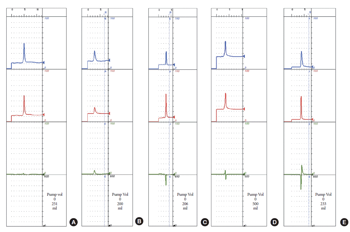
Different spikes in Pdet during the initial cough are illustrated in Fig. 3. Three basic types were identified. Type I showed a minimal change in Pdet. Type II exhibited a monophasic spike during the cough, which could be a positive spike (type IIa) or a negative spike (type IIb). Type III contained a biphasic spike, which could be a positive-to-negative biphasic spike (type IIIa) or a negative-to-positive biphasic spike (type IIIb).
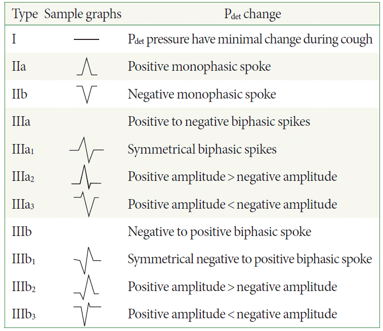
Some biphasic (type III) cough spikes were asymmetric around the baseline. We compared the spike amplitude of each side around the baseline and classified them into 3 subtypes. Types IIIa1 and IIIb1 exhibited equal amplitudes in a biphasic spike (i.e., a symmetric biphasic spike). Types IIIa2 and IIIb2 displayed a biphasic spike with a greater positive amplitude and a smaller negative amplitude. Finally, types IIIa3 and IIIb3 exhibited a biphasic spike with a smaller positive amplitude and a greater negative amplitude. Sample graphs of each type are presented in Fig. 4.
We also investigated the distribution of high quality tracings across these types of initial cough spikes (Tables 3). The percentage of high quality tracings was greatest for type I initial cough traces (P<0.01).
DISCUSSION
Initial Cough Pressure Values and TVRs
As abdominal strength differs among individuals, the Pves and Pabd values during the initial cough varied widely. Another reason for the wide range of pressures is that most of the neurogenic bladder patients in our center had a history of spinal cord injury. Abdominal muscle strength decreases in patients with a spinal cord injury above the thoracic level, which leads to decreased Pves and Pabd values during coughing. Some highlevel spinal cord injury patients cannot even cough. We observed that the 95% range of the cough amplitude for Pves and Pabd were 4ŌĆō62 and 3ŌĆō70 cm H2O, respectively, in the supine position and 9ŌĆō95 and 8ŌĆō98 cm H2O, respectively, in the sitting position among neurogenic patients. The lower extent of those ranges is less than the value of 15 cm H2O that was proposed by Hogan et al. [6] as the minimum peak height that should be generated by a cough. If the cough amplitude for Pves and Pabd is lower than 5 cm H2O, which is classified as a minor dynamic change [3], the signal transmission cannot be fully tested. In this situation, the signal transmission can instead be tested by pressing the abdomen slightly above the superior edge of the symphysis pubis. We recommend the mean cough amplitude in supine position (21 cm H2O) as the proper pressure to apply on the abdomen.
The cough amplitudes were higher in sitting position. This was likely due to the presence of greater pressure loads on the pelvic organs in upright positions. As the influence of abdominal strength and position was eliminated by subtracting Pabd from Pves, the range of cough Pdet values was narrower than that of Pves and was similar in the supine and sitting positions.
We also observed that 95% of the initial resting Pves and Pabd values were between 4 and 30 cm H2O. This range can be used as a reference level for neurogenic bladder patients. This initial resting level is lower than has been observed for nonneurogenic groups [3,7,8]. Whether the difference between different patient groups is statistically significant should be the subject of future studies based on the data obtained at our center.
Initial Cough Spikes and TSPs for Pdet
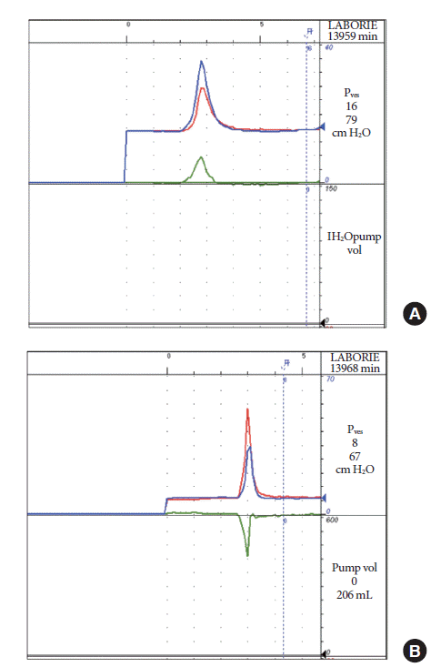
The initial cough is the cough generated before bladder filling. The bladder is empty at that time. When a cough occurs, the pressure inside the bladder (Pves) and around the bladder (Pabd) should exhibit a synchronous change; that is, they should start rising at the same time, peak at the same time, and return to the resting pressure at the same time. In this situation, Pdet should not change, as it is obtained by subtracting Pabd from Pves (Fig. 5A), and the amplitude of minor dynamic changes caused by breathing, talking, and moving is less than 5 cm H2O [3]. In Pdet traces, small spikes corresponding to a pressure change of less than 5 cm H2O are not visually noticeable, so we defined 5 cm H2O as the threshold level for considering pressure changes negligible. All cough spikes with a Pdet change of less than 5 cm H2O can be classified as type I, which reflects the judgment that an amplitude difference of less than 5 cm H2O in Pves and Pabd during a cough can be considered acceptable for good urodynamic practice.
After merging the Pves, Pabd, and Pdet traces into a single channel and amplifying the signals, we noted that when a minor delay took place in pressure transmission, Pdet displayed a symmetric biphasic spike (Fig. 5B). Symmetric biphasic spikes are artifacts that are easy to correct in retrospective studies according to the GUP guidelines. If the transmission delay occurred in Pabd, a type IIIa1 pattern (a positive-to-negative biphasic spike) resulted, whereas a delay in Pves resulted in a type IIIb1 trace (a negative-to-positive biphasic spike). As the double-lumen transurethral catheter has a smaller diameter than the balloon catheter, transmission delays can easily happen in Pves. If the delay happens in the transmission of Pabd, bubbles or blocks in the balloon catheter or tubing might be the problem. Insufficient transmission would also result in a smaller amplitude in Pabd during coughing; if the cough amplitude difference between Pves and Pabd is less than 5 cm H2O, the delay can be considered acceptable.
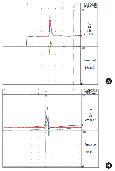
Insufficient transmission can lead to unequal changes in Pves and Pabd during coughing (Fig. 6). An uneven height (height difference >5 cm H2O) in Pves and Pabd during the cough can result in either a positive or negative spike. A positive spike (type IIa) results from a low Pabd during coughing, whereas a negative spike (type IIb) is due to a low Pves during coughing. Low pressure during coughing may be caused by air bubbles in the catheter or tubing. Coughs generate transient high pressure and cause rapidly changing signals. Air in bubbles is compressible and attenuates rapidly changing pressure waves during the transmission process [9]. Other factors can also result in low pressure readings, such as leakage in the system between the transducer and patient, the catheter tip pressing the bladder mucosa, the side hole of the pressure transmission being blocked by mucus or stones, and kinked or blocked tubing. We suggest that incorrect pressure readings due to catheter dislocation are unlikely to be the reason for monophasic cough spikes, because if the catheters become dislocated during coughing, the pressure will not return to the initial resting level.
As shown in Fig. 7, if low pressures are combined with a transmission delay, asymmetric biphasic spikes appear. A low and delayed Pabd value results in a positive-to-negative biphasic spike with a greater positive amplitude (type IIIa2). A low Pabd value and a delayed Pves value result in a negative-to-positive biphasic spike with a greater positive amplitude (type IIIb2). A low Pves value and a delayed Pabd value result in a positive-to-negative biphasic spike with a smaller positive amplitude (type IIIa3). A low and delayed Pves value results in a negative-to-positive biphasic spike with a smaller positive amplitude (type IIIb3).
When monophasic spikes or asymmetric biphasic spikes appear during the initial cough, it alerts the operators that they need to determine the potential reasons for incorrect pressures and carry out a thorough troubleshooting process before filling. Flushing the tubing and catheters, adjusting the position of the catheters, checking the continuity of the tubing, and reconfirming that the catheters are free of bubbles are possible remedial actions.
Our statistical analysis indicated that the percentage of high-quality traces was highest for type I initial coughs (P<0.01). This suggests that if this type of cough signal is obtained at the beginning of the examination, the possibility of obtaining a high-quality test is greater. Therefore, we recommend the presence of a type I cough signal before bladder filling as a reasonable standard.
While we did not analyze coughs during bladder filling, at the end of filling, or before and after voiding in this study, it is likely that the acceptable patterns of cough spikes may be the same. Good cough signals at the end of filling and before and after voiding are also important. Cough signals change during urodynamic testing and we should ensure that most of them are high-quality. If monophasic or asymmetric cough spikes occur continuously, the test should be stopped for trouble-shooting using the remedial actions presented above.
In conclusion, we established TVRs for the initial cough test in neurogenic bladder patients that can be used for quantitative quality control. TSPs for the initial cough signal were described, and we recommend the presence of a good initial cough signal for quality control in urodynamic examinations.