Improved Detection of Urolithiasis Using High-Resolution Computed Tomography Images by a Vision Transformer Model
Article information

Abstract
Purpose
Urinary stones cause lateral abdominal pain and are a prevalent condition among younger age groups. The diagnosis typically involves assessing symptoms, conducting physical examinations, performing urine tests, and utilizing radiological imaging. Artificial intelligence models have demonstrated remarkable capabilities in detecting stones. However, due to insufficient datasets, the performance of these models has not reached a level suitable for practical application. Consequently, this study introduces a vision transformer (ViT)-based pipeline for detecting urinary stones, using computed tomography images with augmentation.
Methods
The super-resolution convolutional neural network (SRCNN) model was employed to enhance the resolution of a given dataset, followed by data augmentation using CycleGAN. Subsequently, the ViT model facilitated the detection and classification of urinary tract stones. The model’s performance was evaluated using accuracy, precision, and recall as metrics.
Results
The deep learning model based on ViT showed superior performance compared to other existing models. Furthermore, the performance increased with the size of the backbone model.
Conclusions
The study proposes a way to utilize medical data to improve the diagnosis of urinary tract stones. SRCNN was used for data preprocessing to enhance resolution, while CycleGAN was utilized for data augmentation. The ViT model was utilized for stone detection, and its performance was validated through metrics such as accuracy, sensitivity, specificity, and the F1 score. It is anticipated that this research will aid in the early diagnosis and treatment of urinary tract stones, thereby improving the efficiency of medical personnel.
INTRODUCTION
Urinary stones can cause lateral abdominal pain, such as side pain, and present in an intermittent pattern, temporarily subsiding before reappearing. In men, the pain may radiate to the lower abdomen, testicle, and scrotum, while in women, it may extend to the vagina. When stones are located near the bladder, symptoms of bladder irritation, such as frequent urination, may also occur. Severe pain can be accompanied by nausea, vomiting, and abdominal swelling, and urinary stones may also cause hematuria [1-3]. The primary cause of urinary tract stones is reduced water intake. This decrease leads to urinary crystals remaining in the urine for extended periods, increasing the likelihood of stone formation. Urinary tract stones are more common in men, occurring at a rate three times higher than in women, and are often seen in individuals in their 20s to 40s. The prevalence of this condition is particularly high in mountainous, desert, and tropical regions, suggesting that diet, temperature, and humidity may contribute to its development. Temperature and season are significant factors in the formation of urinary tract stones; during summer, concentrated urine from excessive sweating can facilitate stone production. Additionally, excessive sun exposure can lead to an overproduction of vitamin D, and a high intake of animal protein can increase the risk of urinary stones by raising the excretion of secondary calcium, phosphates, and uric acid [4,5].
Urinary tract stones can be diagnosed through the identification of the patient’s symptoms, physical examination, urinalysis, or radiographic imaging. Patients with suspected ureteral stones often experience severe pain in the back, and when a urinary tract infection is present, urinalysis may reveal an elevated white blood cell count in the urine. It is important to note that while a simple radiographic scan can confirm the presence of a stone, it cannot diagnose stones that are not radiopaque. Furthermore, ureteral stones may be obscured by pelvic bones or masked by fecal matter or other organs, complicating their detection. Therefore, to ascertain the presence of ureteral stones, hospitals may utilize angiography or computed tomography (CT) [6].
In the field of deep learning vision, high-performing technologies such as the convolutional neural network (CNN) [7] and transfer learning have been developed, along with the vision transformer (ViT) model [8], which has set new state-of-the-art records in many areas. Numerous studies employing artificial intelligence have been conducted in the medical and healthcare fields. Research has been carried out to detect kidney or urinary stones [9-12], manage urinary tract stones [13], predict the risk of urinary tract stones [14], and enhance the resolution of CT images using CNN [15]. However, the ViT model tends to underperform when fine-tuning with limited datasets. Therefore, in this paper, we propose a pipeline that utilizes medical data to determine the presence or absence of urinary tract stones with the ViT model.
MATERIALS AND METHODS
In this study, a total of 150 images were utilized, comprising 100 images of urinary stones and 50 images not containing urinary stones. These images were obtained from patients who visited Sejong Chungnam University Hospital and were verified by medical experts. The collected data were anonymized and, for research purposes, stored on a separate storage device in DICOM format. Fig. 1 represents the CT scans used in the research.
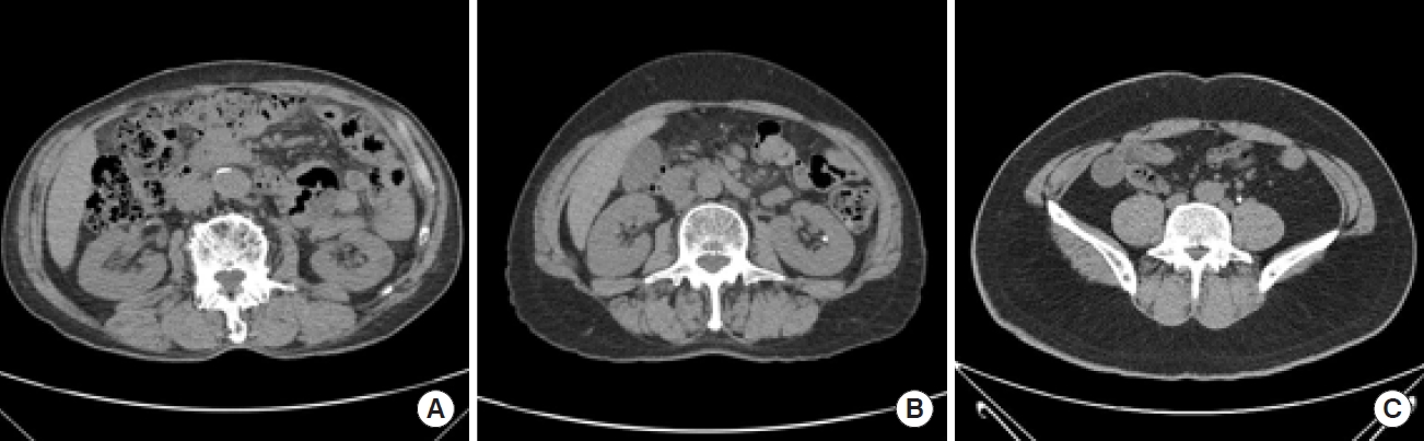
Sample images from the dataset: normal (A), stone (B), and ureter stone (C).
For stone detection, the ViT backbone model was utilized, necessitating the resizing of input images to 224×224 dimensions. A notable feature of the ViT model is its capacity to achieve outstanding performance when fine-tuned with high-resolution images, irrespective of the resolution of pre-trained images. To facilitate this, the technique of histogram smoothing [16] is applied. Histogram smoothing modifies the pixel value distribution within the image to create a more uniform distribution, which in turn enhances image contrast and improves performance.
Furthermore, extensive research has been conducted in the field of medical CT imaging, involving super-resolution techniques to enhance image resolution. Building on this research, the super-resolution CNN (SRCNN) [17] was employed in this experiment to increase the resolution of the data used. SRCNN is a technology that upscales low-resolution images to high resolution, restoring fine details and enhancing the overall image resolution. This improvement in data quality contributes to the enhancement of the model’s performance. The SRCNN employs bicubic interpolation to upscale a single low-resolution image to the desired size. It transforms the target low-resolution image, denoted as Y, into F(Y), with the goal of identifying a mapping function F that enables F(Y) to closely approximate the original high-resolution image X. The process adheres to the following steps:
F1(Y)=max(0,W1×Y+B1) (1)
F1(Y)=max(0,W2×F1(Y)+B2) (2)
F(Y)=W3×F2(Y)+B3 (3)
First, patch extraction and representation: This process involves extracting patches from the low-resolution image Wai using the first layer of a CNN and representing each patch as a high-level vector [1]. Next, nonlinear mapping: The high-dimensional vectors generated in the process [1] are nonlinearly mapped to other high-dimensional vectors. Finally, reconstruction: The high-resolution representations produced through the aforementioned processes are amalgamated to construct the final high-resolution image. At this stage, the resulting value should closely approximate the actual correct answer, X.
Data Augmentation
High-quality data play an important role in improving model performance. Therefore, extensive research has focused on data augmentation. This field has progressed from elementary techniques, such as random horizontal or vertical rotations and shifts of images, to more sophisticated methods including aspect ratio adjustments, image flipping, and the use of generative models.
Among them, CycleGAN [18] is a powerful model used to convert images into different styles within the framework of generative adversarial networks. The key idea behind this model is to effectively transform data from one domain to another while maintaining consistency with the original image. Cycle-GAN has been employed in various fields, including art, style transformation, photo coloring, and landscape transformation, significantly aiding in the creation of images with diverse styles and enhancing data diversity.
Vision Transformer
In this experiment, the ViT model played a crucial role in addressing the limitations of the existing CNN architecture regarding spatial structures. ViT segments images into small patches and converts these patches into vectors to represent the spatial structure of the images. This method relies on the attention mechanism and transformer architecture, facilitating efficient learning of both global and local features.
The advantage of ViT lies in its ability to learn efficiently while preserving the immutability of images. Consequently, this paper introduces a model designed to detect the presence or absence of urinary stones by effectively learning the spatial structure of CT images. The model identifies urinary tract stones by taking into account the spatial structure of the image, and Fig. 2 illustrates the architecture of this model. This approach allows ViT models to be utilized in experiments for efficient learning and to enhance the performance of urinary tract stone classification.
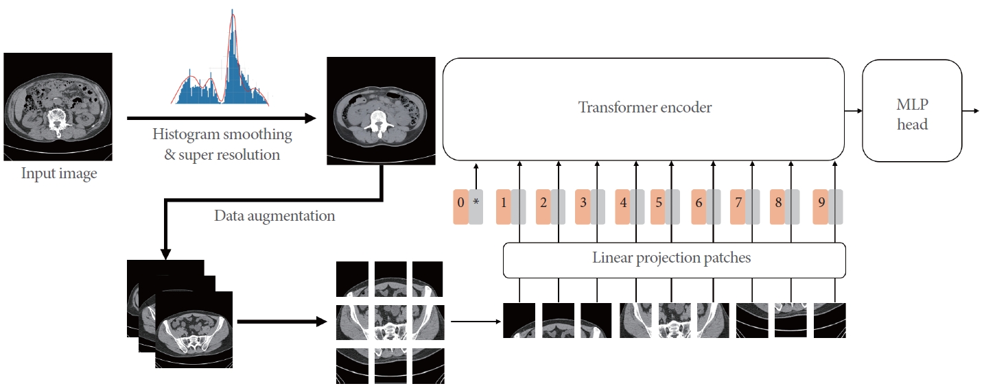
Model architecture. MLP, multilayer perceptron.
Model Evaluation
In this study, a 5-fold cross-validation method was employed to assess the model’s performance. Cross-validation is a technique used to evaluate the performance and generalization capabilities of machine learning models. It is particularly effective for small datasets, providing a more accurate evaluation than a single training-test split. The process involves dividing the entire dataset into k subsets (folds) and using each fold in turn for training and testing, ensuring that every data point is included in the test set at least once.
In this process, the model’s performance is measured on each fold, and the average of these results serves as the final performance indicator. The study employed accuracy, precision, and recall scores as performance evaluation metrics to assess the model’s effectiveness. Accuracy denotes the proportion of correct predictions out of all predictions made, precision indicates the proportion of positive predictions that are correct, and recall reflects the proportion of actual positive instances that were correctly identified. Furthermore, the diagnostic performance of the model in this study was validated using this method.
RESULTS
A pipeline for detecting the presence and absence of urinary stones was developed using a ViT model. To evaluate the model’s performance, accuracy, precision, and recall were used as indicators. Additionally, to ensure the model’s generalizability, 5-fold cross-validation was employed. Table 1 presents the results of the cross-validation comparison among different deep learning and machine learning models.
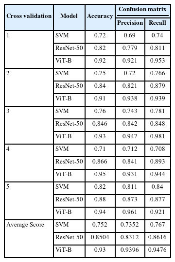
Comparison of model performance
DISCUSSION
Urinary tract stones cause lateral pain and various symptoms, and they occur with particularly high frequency in men and in young people in their 20s to 40s. The primary cause is an increase in urine concentration due to insufficient water intake. Additionally, environmental factors play a role, particularly in highland, desert, and tropical regions. Given these factors, the absence of urinary tract stones is regarded as a significant medical issue. The diagnosis of urinary tract stones may involve evaluating the patient’s symptoms, conducting a physical examination, performing a urine test, or utilizing imaging techniques. However, standard radiography may not always detect all stones, occasionally necessitating more advanced imaging methods such as angiography or CT scans.
This study introduces a pipeline designed to leverage medical data for the diagnosis of urinary tract stones. The dataset employed in this research comprised 150 CT images sourced from Sejong Chungnam National University Hospital. These included 100 images from patients diagnosed with urinary tract stones and 50 images from patients without urinary tract stones.
In the data preprocessing phase, the SRCNN model was utilized to enhance image resolution. This preprocessing step enhances the model’s performance and facilitates learning that takes into account the spatial structure.
Data augmentation plays an important role in improving the performance of the model, particularly through the application of technologies like CycleGAN to increase data diversity. Such improvements bolster the models’ generalization capabilities, allowing them to leverage a wide array of data styles.
The ViT model learns spatial structure by segmenting images into patches and transforming them into vectors, thereby enabling efficient learning while preserving the images’ immutability. Utilizing this model, we developed a system for detecting urinary tract stones, which demonstrated effective performance in our experimental results.
A 5-fold cross-verification method was used to evaluate the model’s performance, and accuracy, precision and recall scores were used as evaluation indicators. The results yielded a precision of 0.9396, a recall of 0.9476, and an accuracy of 0.93. These outcomes are anticipated to validate the model’s diagnostic capabilities and aid in the early diagnosis and treatment of patients with urinary tract stones.
Notes
Conflict of Interest
No potential conflict of interest relevant to this article was reported.
AUTHOR CONTRIBUTION STATEMENT
· Conceptualization: TKW
· Data curation: HSC
· Formal analysis: JSK
· Methodology: TKW
· Project administration: SJE
· Visualization: SJE
· Writing - original draft: HSC
· Writing - review & editing: JSK